
By Roxanne Sicklen, clinical specialist sonographer, Royal Free London NHS Foundation Trust and senior teaching fellow, King's College London
Ectopic pregnancies within the uterus disproportionately contribute to maternal morbidity and mortality in the first trimester.
Pregnancies located within a previous caesarean section scar can be missed if the ultrasound practitioner is not alert to the possibility of this condition arising and fails to perform a systematic assessment.
With NHS Maternity Statistics, 2024-25 reporting a 45 per cent caesarean section rate in England1, the incidence of caesarean scar ectopic pregnancy (CSP) is expected to rise in parallel.
This CPD article outlines key considerations for ultrasound practitioners to support accurate diagnosis of CSP.
Six key points
1. History taking is essential
A thorough clinical history is the foundation of accurate interpretation. You cannot be alert to the possibility of identifying a caesarean scar pregnancy if you don’t know the patient has had a previous caesarean section.
2. Types of caesarean scar pregnancy
According to European Society of Human Reproduction and Embryology terminology2, CSPs can be classified as complete or partial, based on the relationship with the endometrial cavity:
- Complete CSP: The gestational sac is entirely embedded within the myometrial defect of the caesarean scar, with none of the sac in the endometrial cavity.
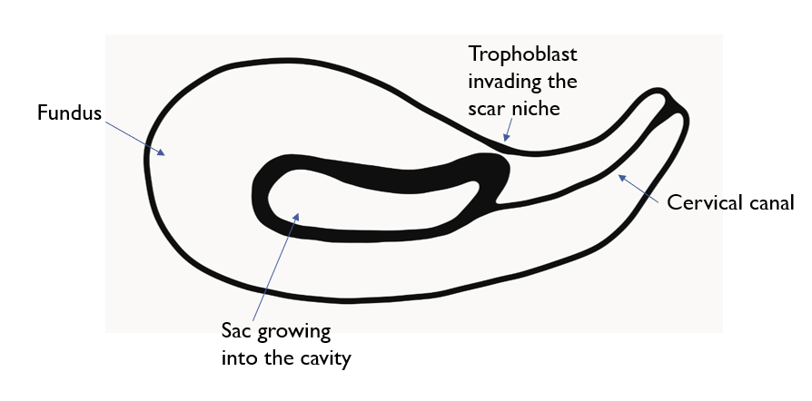
- Partial CSP: The gestational sac lies partly within the endometrial cavity but has part of the sac or trophoblast in the scar niche. These are more subtle and may be mistaken for low-lying, normally sited (eutopic) pregnancies.
Figure 1: Complete caesarean scar ectopic pregnancy
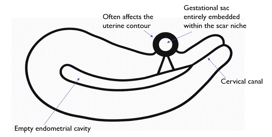
Figure 2: Partial caesarean scar ectopic pregnancy
3. Sagittal ‘sac to cervix’ view
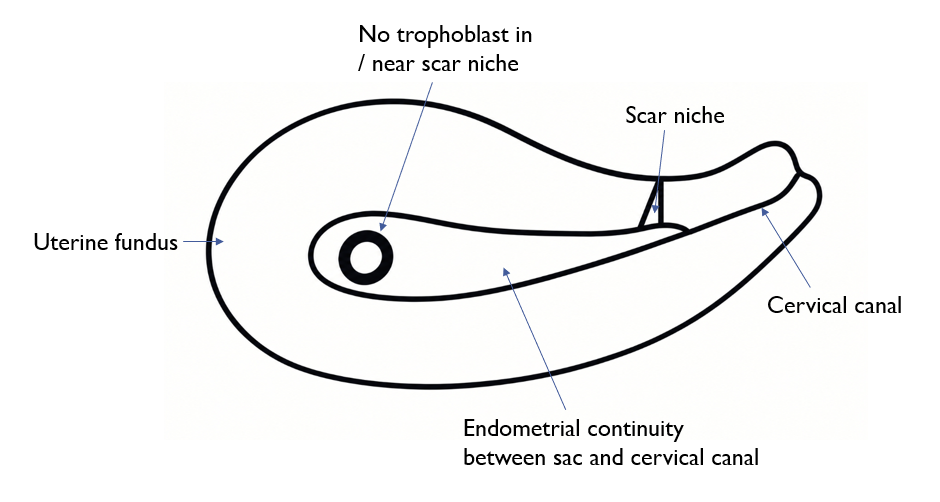
A correctly sited (eutopic) pregnancy should lie entirely within the endometrial cavity. Every first-trimester scan should include a midline sagittal assessment and an archived image of the whole uterus, demonstrating continuity between the cervical canal and the gestational sac. The relationship between the sac and any previous caesarean scar site should also be examined.
Figure 3: Sagittal ‘sac to cervix’ view
4. Trophoblast
Confidently identifying the implantation site is key to diagnosing caesarean scar pregnancies. In partial CSP, the bulk of the sac may appear within the uterine cavity, but careful evaluation reveals the echogenic trophoblast extending into the caesarean scar niche. If undetected, excessive blood loss during management of miscarriage or complications from subsequent placenta accreta spectrum are common.
5. Doppler
Colour or power Doppler is a useful tool to assess for trophoblastic vascularity in the scar niche to differentiate partial CSP from a normally sited (eutopic) pregnancy that has implanted posteriorly in the lower cavity.
Any Doppler use should be judicious, ensuring that diagnostic benefit outweighs any theoretical risks associated with higher ultrasound power settings in early pregnancy.
6. Second opinions
Where uncertainty exists about the location of a pregnancy, a second opinion should be sought from a second operator. Many units will have senior decision makers available, but the option to refer the patient to a specialist tertiary unit also exists. These units have expertise in complex early pregnancy cases and can provide definitive diagnosis and management planning if required.
Reflection prompts
- What is your usual process for a first-trimester scan (including nuchal / dating scans)? Do you routinely assess and image the pregnancy location within the uterus?
- Do you take a history from pregnant women/people to understand their individual risk factors and what changes might you make in the future?
- Are you aware of the link between a history of previous caesarean section and the increased risk of placenta accreta spectrum?
- Does your department have a protocol to risk-stratify patients?
- Are there any changes you can make to your practice to increase the detection of CSP, in light of this CPD article?
References
- NHS England (2025). NHS Maternity Statistics, 2024–25. https://digital.nhs.uk/data-and-information/publications/statistical/nhs-maternity-statistics/2024-25
- The ESHRE working group on Ectopic Pregnancy, Kirk E, Ankum P, Jakab A, Le Clef N, Ludwin A, Small R, Tellum T, Töyli M, Van den Bosch T, Jurkovic D. (2020). Terminology for describing normally sited and ectopic pregnancies on ultrasound: ESHRE recommendations for good practice, Human Reproduction Open, 2020(4); hoaa055. https://doi.org/10.1093/hropen/hoaa055