
Written by Angie White, senior lecturer at Brunel University, sonographer and vascular technologist
This CPD article aims to provide an overview of how to recognise Pelvic Venous Disorders (PeVD), an underdiagnosed cause of chronic pelvic pain.
By recognising the symptoms of PeVD in patients, and the differences between those assigned male and assigned female at birth, sonographers can ensure accurate diagnosis and guide timely clinical management.
This article outlines six key points followed by six reflection prompts, to enhance your expertise in diagnosing PeVD using ultrasound imaging.
What is PeVD?
Pelvic Venous Disorder (PeVD) is a broad term used to describe an abnormality of the veins within the pelvis (Meisner et al., 2021). These abnormalities occur due to venous reflux, where retrograde blood flow pools in the pelvic veins, increasing venous pressure leading to vein dilatation and formation of varicose veins.
Venous reflux is due to absent or partially functioning valves, most commonly in the gonadal (testicular/ovarian) and internal iliac veins and their tributaries. Less commonly PeVD can result from venous compression, obstruction or congenital vascular anomalies.
In those assigned male at birth, PeVD is well documented and reported on imaging tests, most typically in the form of testicular varicocoele. Unfortunately the same standard does not apply for those assigned female at birth. In females, ultrasound can identify these abnormal veins and assess for reflux, making sonographers key contributors to the diagnosis of PeVD. An awareness of the clinical presentations and ultrasound findings is therefore essential.
Clinical Presentations
1. Presenting symptoms
Some people with PeVD are asymptomatic.
When symptomatic, PeVD symptoms (commonly known as Pelvic Congestion Syndrome) can include (* denotes symptoms that are unique):
- Chronic pelvic pain (CPP+), and a feeling of heaviness or ‘dragging’ in the pelvis*
- Lower back pain, worsening over the course of the day to the point where it is only alleviated by lying down (pain typically resolves over-night)*
- Pain worsening with prolonged standing*
- Irritable bladder or urinary frequency/urge incontinence
- Irritable bowel symptoms, especially bloating
- Itching, throbbing or heavy dragging sensation in the vulvar/labial area
- Leg symptoms (aching/heavy upper thighs and/or posterior thigh pain with prolonged sitting
- Deep dyspareunia (pain may continue after intercourse, sometimes lasting for days)*
- Symptoms are persistent (non-cyclical), however can worsen with cycle (most often premenstrual, but also with menses and during ovulation)*
CPP is defined by the Royal College of Obstetricians and Gynaecologists (RCOG) as pain in the lower abdomen or pelvis lasting longer than six months (RCOG, 2012). CPP due to PeVD is often referred to as Chronic Pelvic Pain of Venous Origin (CPP-VO) or Pelvic Congestion Syndrome (PCS).
2. Presenting signs
Most often varicose veins (varices) are seen, whether noted internally or externally. Specific sites may be:
Internal:
- Renal varices
- Pelvic varices (adnexal/periuterine/arcuate veins of the uterus)
External:
- Vulvar varices (which are typically reported during pregnancy)
- Haemorrhoids (whether internal or external)
- Lower extremity varicosities that originate from the pelvis (typically noted in the upper thigh/groin area); recurrence in common after treatment if there is a connection to the pelvis
PeVD is often overlooked
PeVD is thought to contribute to up to 30% of cases of CPP, yet remains under-recognised and underdiagnosed. This can delay appropriate treatment and increase the impact on patients and healthcare systems (Ni et al., 2026).
Diagnosis of PeVD with ultrasound
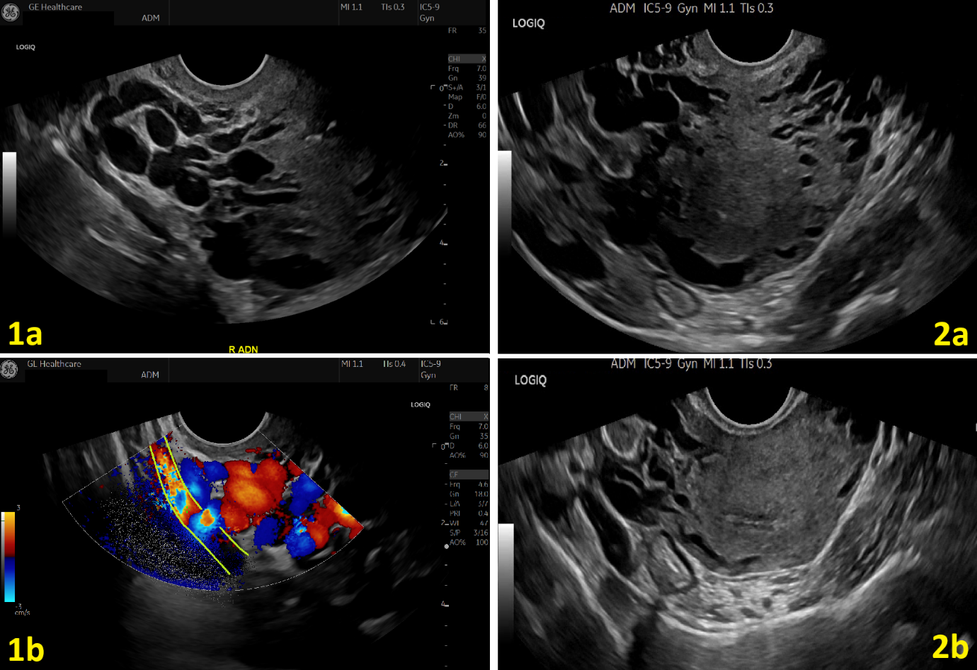
Figure 1. Transvaginal ultrasound appearances of pelvic venous dilatation of PeVD. 1a) Grey-scale image demonstrating multiple dilated, tortuous pariuterine and adnexal varicosities. 1b) Colour Doppler imaging confirms venous flow within the dilated vessels, with venous reflux elicited during the Valsalva manoeuvre of the internal iliac vein (delineated with green lines/green cross). 2a) Grey-scale image obtained with minimal transducer pressure, demonstrating prominent dilated arcuate and adnexal veins. 2b) Repeat image in the same patient with moderate transducer pressure, demonstrating compressibility and partial collapse of the pelvic veins, consistent with a venous origin.
Signs of PeVD in those assigned female at birth can be seen sonographically. Typical ultrasound findings include dilated arcuate veins within the myometrium, and dilated, tortuous (varicose) veins noted within the adnexae (Figure 1- 1a). Gentle probe pressure should be used to avoid compressing the veins and potentially obscuring findings (Ni et al., 2026) (Figure 1- 2a/2b). Colour Doppler with a controlled Valsalva manueovre can be used to delineate varicosities from other pathologies. A low PRF should be utilised to observe the slow flow in the vessels (Figure 1- 1b).
There is no universally accepted vein diameter threshold for diagnosing PeVD in females. Vein diameter does not always correlate with symptoms or reflux, for example nulliparous women often have smaller diameter pelvic veins (dos Santos et al., 2015). A diagnosis should consider both the ultrasound findings and patient clinical presentation. It is therefore essential for the sonographer to explore symptoms with the patient.
Reporting the findings
Sonographers should report pelvic varices, when identified, and document if the patient reports symptoms that might indicate PeVD. Asymptomatic PeVD should be reported, as the patient may develop future symptoms, or may present with recurrent and previously untreatable varicose veins of the legs.
Example reporting phrases that could be used are:
- “Dilated adnexal and arcuate veins noted, however patient reports no symptoms of pelvic venous disease.”
- “Dilated adnexal veins noted bilaterally, patient reports symptoms that correlate with pelvic congestion syndrome.”
Treatment
Appropriate recognition and reporting can support referral, diagnosis of underlying pathology, and treatment. The most common treatment is coil embolization of the affected gonadal and/or internal iliac veins. Reported treatment outcomes are positive, with efficiencies in reducing CPP (de Carvalho et al., 2023).
Reflection prompts:
- How confident are you in recognising the ultrasound features of PeVD?
- Do you routinely ask patients about symptoms that might be associated with PeVD?
- How might a patient describe their pain if they present with chronic pelvic pain associated with PeVD?
- How might PeVD present differently from other causes of chronic pelvic pain?
- Does reporting of varices /varicoceles differ in males and females? (For example, if you identified a varicocele during a testicular ultrasound, would you include this in the report? How would you approach a similar finding on a transvaginal ultrasound scan currently? If your reporting approach differs between the two examinations, what factors influence that decision?)
- What is your local patient pathway for PeVD (for both diagnosis and treatment)?
References:
- de Carvalho SFC, Metzger PB, Fernandez MG, Ribeiro WB, Nogueira AKS and Souza, JPRe. (2023). Pelvic venous reflux embolization in the treatment of symptomatic pelvic congestive syndrome: A systematic review with meta-analysis. Journal of Vascular Surgery: Venous and Lymphatic Disorders, 11(2), 412-421.e6. https://doi.org/10.1016/j.jvsv.2022.10.005
- Dos Santos SJ, Holdstock JM, Harrison CC, Lopez AJ, Whiteley MS. (2015) Ovarian Vein Diameter Cannot Be Used as an Indicator of Ovarian Venous Reflux. Eur J Vasc Endovasc Surg. 49(1):90-4. doi: 10.1016/j.ejvs.2014.10.013.
- Meissner MH, Khilnani NM, Labropoulos N, et al. (2021). The Symptoms-Varices-Pathophysiology classification of pelvic venous disorders: A report of the American Vein & Lymphatic Society International Working Group on Pelvic Venous Disorders.J Vasc Surg Venous Lymphat Disord. 1(9): 568–584. https://doi.org/10.1016/j.jvsv.2020.12.084
- Ni T, Friedman I, Smith J, Liu B, Melchior M and Brown A D. (2026). Pelvic venous disorders in women: Diagnosis and management for gynecologists. International Journal of Gynecology & Obstetrics. https://doi.org/10.1002/ijgo.70955
- Royal College of Obstetricians and Gynaecologists (RCOG) (2012) The initial management of chronic pelvic pain. Green-top Guideline No. 41. London: RCOG.