
Since the publication of saving babies lives care bundle (SBLCB) version 2, demand for uterine artery Doppler screening for fetal growth restriction (FGR) has grown. It continues to be the case in SBLCB version 3.
Do you ever feel like it is impossible to sample the uterine arteries?
Below are some tips to help you master the art of finding the uterine artery.
Six Key Points
- The best way to find the uterine artery is to start in longitudinal section just above the pubic bone. Then move laterally and rotate the probe slightly to elongate the external iliac vessels.
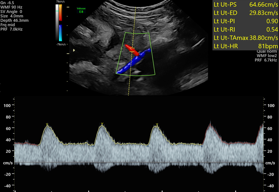
- A large colour box will help you to identify the uterine artery crossing the external iliac vessels. Before sampling you can then reduce the size of the box.
- Dip your probe to improve the Doppler angle to bring the uterine artery closer to vertical and thus obtain the best possible trace. Increase your pulse repetition frequency (PRF) to remove aliasing before applying the pulse wave.
- Optimise your spectral trace by adjusting the sweep speed and baseline to ensure you can trace at least 3 complete waveforms. Auto-trace can then be applied, and Doppler sensitivity altered to ensure the peaks and troughs of the waveforms are correctly identified.
- Each uterine artery should be sampled at least 3 times, 1.5cm downstream (anterior to the external iliac vessels). The lowest PI should be reported if all the traces are of equal quality.
- Perseverance is the key to successful assessment of the uterine arteries.
Reflection Prompts:
- Do you know how to use your machine settings to optimise your Doppler image? How do you change the PRF, sweep speed, baseline, sensitivity and Doppler gain?
- Revisit your vascular anatomy - do you understand where to sample the uterine artery downstream of the crossover and where the uterine artery arises from the internal iliac artery?
Further Reading:
Bhide, A et al. (2021), ISUOG Practice Guidelines (updated): use of Doppler velocimetry in obstetrics. Ultrasound Obstet Gynecol, 58: 331-339. https://doi.org/10.1002/uog.
Elearning for healthcare. My elearning - Clinical Imaging - 21 Ultrasound - Obstetric - e-IRI_21_05 - Ultrasound: Saving Babies Lives