Prostate Cancer UK (PCUK) and the SoR have produced a base set of protocols for mpMRI, an imaging technique that allows 25 per cent of men with suspected prostate cancer to avoid an invasive biopsy procedure.
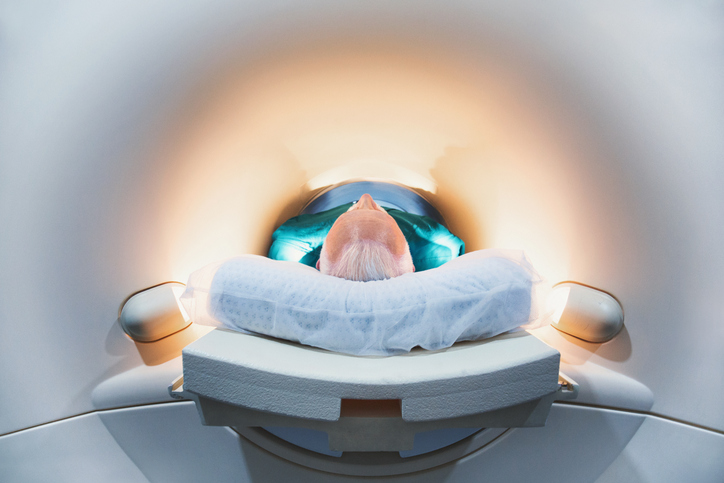
Multiparametric magnetic resonance imaging (mpMRI) is now widely recognised as the frontline method of diagnosing prostate cancer, but for mpMRI to be effective, the images captured need to be of very high quality for accurate, unambiguous interpretation. NICE guidelines recommend that mpMRI be conducted before biopsy.
PCUK has partnered with the Society to produce a set of protocols for the technique.
Darren Walls, the author of the protocols and a research radiographer at University College Hospital, London, said: “This imaging guidance is important as it provides a framework for MR departments to optimise scanners to produce high quality images of the prostate gland for diagnosis and local staging of prostate cancer.”
The protocols employ the imaging techniques used in the prostate imaging study trial. The multiparametric package includes small field of view T2 images, diffusion weighted imaging and dynamic contrast enhanced T1 images of the prostate. This combination is specified by recent UK clinical consensus and its wide adoption means that wherever in the UK a patient has a mpMRI prostate scan, there will be consistencies in the technique and the reporting.
A further benefit of a unified approach to mpMRI is the possibility for a bank of images to be collated, to provide training and feedback for radiographers new to the technique.
Darren said, “(This guidance) provides the opportunity to test an imaging protocol, review the images and then act on the feedback. Radiographers will feel more competent in adapting scan parameters to avoid/reduce artefacts in mpMRI. It will develop the MR radiographer’s knowledge of the underpinning physical principles employed in MRI.”
The mpMRI protocol document can be downloaded from Prostate Cancer UK.
mpMRI before biopsy: Improving diagnosis for men with prostate cancer
Q&A with Darren Walls, author of the protocols
Why are the protocols for mpMRI important?
This imaging guidance is important because it provides a framework for UK MR departments to optimise their 1.5Tesla MR scanners to produce high quality images of the prostate gland for diagnosis and local staging of prostate cancer. High quality MR images are crucial to help diagnose and manage men with newly diagnosed prostate cancer. The protocol strives to provide some consensus on the need for small field of view T2 images, diffusion weighted imaging and dynamic contrast enhanced T1 images of the prostate, in light of the recent UK consensus document that has been published. Therefore, wherever in the UK a man has a mpMRI prostate scan there will be similarities in the technique. A similar technique equates to a more streamlined approach for radiological reporting and training of MR radiographers.
Who should be using these protocols?
The imaging protocols should be used by MR departments which wish to commence mpMRI at 1.5T for men with newly diagnosed prostate cancer. Other centres which currently perform the technique may use this protocol to adapt their current mpMRI, so as to integrate some of the recommendations from pertinent journal publications.
What else is necessary for an MRI scan of high enough quality to rule men out of biopsy?
Radiographers will interpret the high quality MR scans, so training is an important issue and there is a need for a bank of mpMRI images to be collated to provide training and feedback for radiographers new to this technique. The scans will then be assessed to decide on appropriate patient management, either to perform a TRUS biopsy or not.
Are there significant differences when using a 3T MRI scanner?
A 3T mpMRI protocol will vary in the range of scan parameters used in a 1.5T system. A higher field strength can be used to bolster image resolution and or/improve upon overall scan time. Hopefully in the future, we may be able to work with centres which currently employ 3T mpMRI sequences and create a second imaging guidance document.
Are these a good basis for training in mpMRI imaging?
Yes, I feel an imaging guidance document is valuable for training MR radiographers to develop their skills in creating imaging protocols bespoke to their MR system. It provides an opportunity to test an imaging protocol, review the images and then act on the feedback. Radiographers will feel more competent in adapting scan parameters to avoid/ reduce artefacts in mpMRI. It will develop the MR radiographer’s knowledge of the underpinning physical principles employed in MRI.