
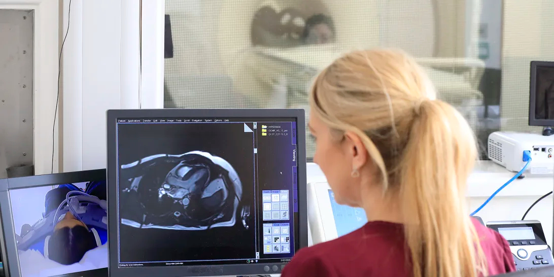
The University of Oxford has successfully used artificial intelligence in place of contrast dye during heart MRI scans.
A study conducted by the University’s Radcliffe Department of Medicine, entitled 'Myocardial Scar Assessment Using Artificial Intelligence-Powered Contrast-Free MRI: A Prospective Multicenter Study of Virtual Native Enhancement', could allow heart MRI patients to avoid using contrast media entirely, reducing discomfort and scan time.
The multicentre, blinded validation published its results this month (15 July) in the Journal of the American College of Cardiology, and is the first of its kind to explore an AI technique called Virtual Native Enhancement (VNE) – which creates 'virtual contrast' heart MRI images without injecting dye into the bloodstream.
A step towards real-world patient care
Professor Qiang Zhang, lead author from the University of Oxford, said: “Many AI tools show promise during technical development, but very few are tested rigorously in real clinical environments. This study shows that AI-powered virtual contrast imaging can work reliably across independent hospitals and clinical teams, which is an important step towards real-world patient care.”
Cardiac MRI typically requires injection of a gadolinium-based contrast agent to allow scanners to pick up scars and other heart damage.
With this AI technique, however, researchers claim the technology could shorten scans, reduce costs, and make cardiac MRI available to more patients. This includes some people with advanced kidney disease, mothers who are pregnant or lactating, and children.
Potentially avoiding contrast altogether
Teams at the University of Leeds and at the Fuwai Hospital of the Chinese Academy of Medical Sciences in China independently assessed scar presence on scans taken using contrast dye on 136 patients, from May of 2023 until March of 2024. The Oxford team then generated VNE images using only precontrast images – and produced scans of similar quality.
Oxford researchers generating the AI images had no access to the original, contrast-enhanced scans or patient clinical information during image generation and analysis. Independent clinicians then assessed the images blindly, reducing the risk of bias.
When the AI-generated images were judged to be of high quality, VNE detected heart attack scars with around 94 per cent accuracy, comparable to conventional contrast-enhanced MRI.
Independent image readers said for around 70 per cent of heart attack patients in the study, AI-generated images were able to accurately mimic the kind obtained after contrast. This means that more than two-thirds of patients could potentially avoid contrast injections altogether, without reducing diagnostic accuracy.
Using AI safely and reliably
While many systems perform well in early studies in single laboratories, their results often fail to make the jump to routine healthcare because of challenges around image quality, reproducibility, or differences between hospitals and scanners.
Professor Stefan Piechnik, joint senior author, added: “That is why this work matters beyond heart imaging alone. It shows how AI can be developed, tested and introduced into healthcare in a safe and reliable way.”
The study is a collaboration between the University of Oxford Radcliffe College of Medicine, the University of Leeds, and Fuwai Hospital of the Chinese Academy of Medical Sciences in China, funded by the British Heart Foundation and Kusuma Trust.
Find out more information about the study, and read the results in full, online here.
(Image: MRI scan, via University of Oxford)