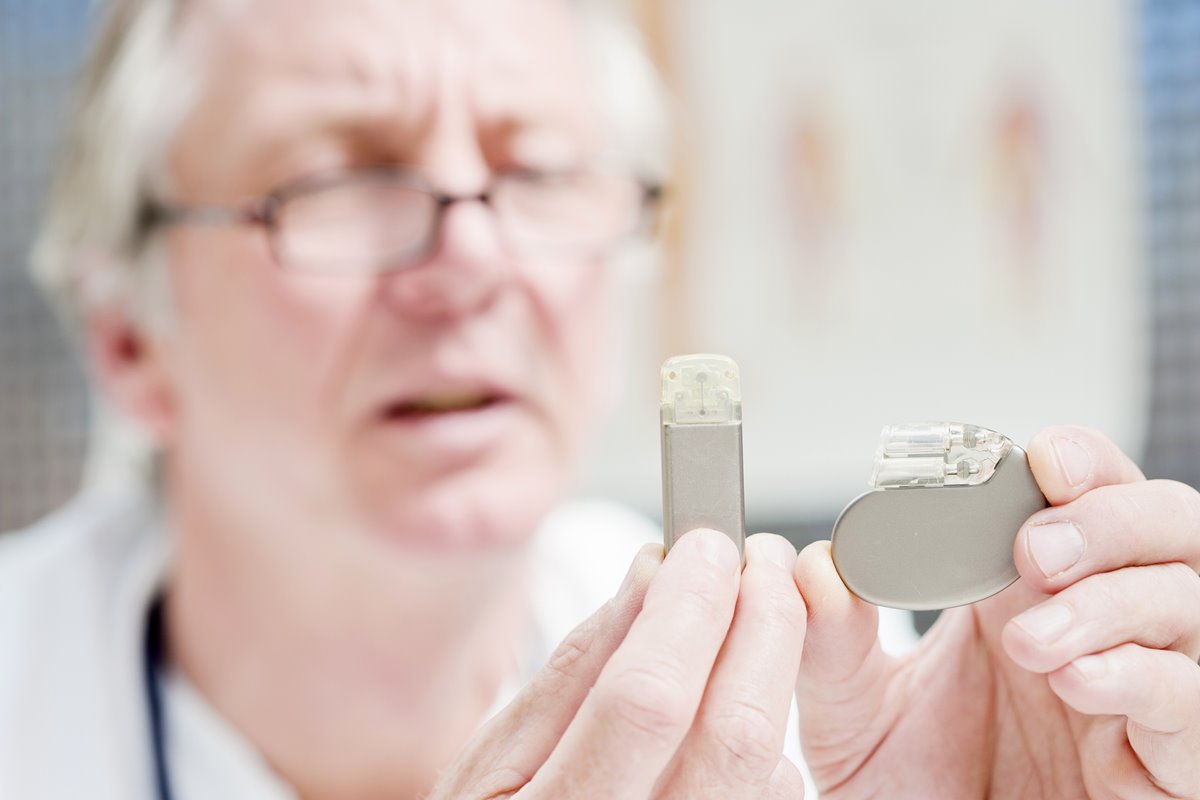
There has been much recent discussion amongst our members and the wider MR community regarding MRI scanning of patients with pacemakers.
A Channel 4 news item broadcast in September 20181 highlighted the lack of access to MRI for many patients with pacemakers. A statement published by the Royal College of Radiologists and the British Cardiovascular Society2, encouraged departments to increase access to MRI for this patient group, although bringing the subject to the forefront seems to have caused some disappointment for our members because it appears there are barriers to providing access for these patients which are not entirely within the control of radiology departments.
Furthermore, confusion is caused by discussing patients with more recent MR Conditional pacemakers alongside patients with older legacy devices, referred to as non-MR Conditional, which historically have been considered a complete contraindication for scanning
To allow MRI pacemaker scanning, a multi-professional approach is required with expertise from cardiology, radiology and MRI physics all coming together to support a successful service.
Aileen Wilson (AW), president-elect of the British Association of MR Radiographers BAMRR; Dr Charlotte Manisty (CM), consultant cardiologist from Barts; Geoff Charles-Edwards (GCE), consultant MR physicist, Guy’s and St Thomas‘; Jonathan Ashmore (JA), MR physicist, NHS Highland; and I explore some of the issues related to scanning patients with pacemakers below.
What is the current situation regarding lack of access to MRI scans for patients with pacemakers?
CM - There are over 400,000 patients in the UK with cardiac pacemakers or implantable cardioverter defibrillators (ICD), and these patients tend to be older, frailer and with more co-morbidities, meaning they have increased demand for MRI compared to the general population. In the UK, estimates suggest there are 50,000 scans a year needed for cardiac device patients, but latest data suggest that only around 1000 scans a year are actually being performed 3. Equity of access would be likely to result in around 1% of adult MRIs being done on pacemaker/ICD patients.
Why is access to MRI for these patients currently so limited?
CM - Prior to manufacturers developing MR Conditional pacemakers in 2008, scanning of these patients did not take place. Manufacturers now produce MR Conditional pacemakers and defibrillators, and in the UK these are now implanted as standard of care. Whilst many departments do scan MR Conditional devices, many still do not, meaning that patients still experience difficulties accessing scans.
AL and AW - Reasons for lack of MRI provision for these patients may include: Lack of access to/or interaction with cardiovascular departments, or no cardiovascular department
- Lack of access to MR physics expertise
- Remote siting of scanners/mobile units
- Low field strength scanner unable to meet diagnostic performance requirements for MRI
- Increased time and cost implications for departments.
JA - From an MR physics perspective, there is at times confusion regarding parameters for scanning pacemakers. Historically, MR Conditional pacemakers have had scan location restrictions,
SAR (specific absorption rate) restrictions, as well as a variety of other limitations such as a complete contraindication for scanning pacemakers with any other metal implant present. Such restrictions seem to be on older systems, but uncertainty around this can often cause departments to be fearful.
What processes should be in place for scanning patients with MR Conditional pacemakers?
AL/AW - In addition to complying with the manufacturers’ conditions for safe operation4, we advise that departments formulate a local policy with reference in the local rules.
Such a policy may include:
- A patient pathway
- Clearly defined roles and responsibilities of the radiology department, the radiographic staff and the cardiovascular department
- A list of any contraindications, potential adverse events and emergency procedures
- A process for obtaining the current manufacturers’ specific operating instructions and any local specific instructions. This should include a mechanism for checking that both the pacemaker and the leads are MR conditional.
JA - A robust standard operating procedure is important with well-defined roles and responsibilities, with the support of senior management and clinical leads. Training is extremely important too for the staff who are directly involved in creating the service. Often this requires them to attend external courses, or visit a department who already has the service in place.
More local training is also extremely useful, particularly to ensure the department in general is comfortable with scanning these devices which were once a complete contraindication.
What are the essential requirements and common MR conditions required for scanning MR Conditional pacemakers? What do radiographers need to know to ensure these are met?
CM - The most important thing is that no pacemakers or defibrillators can be scanned without being re-programmed to MRI mode, even if MRI-conditional. This therefore requires a cardiac physiologist or cardiologist to check and re-program the device before and immediately after scanning. Patients also must be monitored throughout the study, and there must be an immediate life support/advanced life support qualified person available throughout the scan.
JA – Once the device has been put into a ‘safe for MRI’ mode and suitable monitoring is in place, in terms of MRI sequences these can be scanned much the same as say passive conditional implants, ie much like the implants listed on mrisafety.com5).
This essentially means you can scan without changing the scanner settings at all. The pacemaker conditions will typically specify that the scan needs to be undertaken in normal mode though and some scanner vendors do suggest scanning in first level mode as default. Hence, in this case, normal mode should be selected. In some older devices, there are scan exclusion zones or a requirement for reduced SAR.
What about pacemakers that some people call non-MR Conditional, or legacy devices? The MHRA guidelines suggest anything that is not labelled as MR Safe or MR Conditional should be considered as MR Unsafe? How can we even be considering scanning something that is unsafe?
GCE – To answer this, first there needs to be a quick discussion about the international standard for labelling items in the MR Environment. Currently, this has three categories: MR Safe, MR Conditional and MR Unsafe. The definitions of the first two are generally very helpful for MRI units. In both cases the manufacturer is providing assurance that it is safe to take the item into the MR environment, although importantly in the latter case of MR Conditional, this is only when all of the stated conditions are met. (We’ll ignore for the time being that some MR Conditional statements are ambiguous and/or difficult to verify.)
The implication is that everything else falls into the third classification of MR Unsafe, defined as “an item which poses unacceptable risks to the patient, medical staff or other persons within the MR environment.” This definition is more contentious.
Firstly, there is a spectrum of risk associated with bringing items that are not labelled as MR Safe or MR Conditional into the MR environment. For example, a ferromagnetic gas cylinder obviously is associated with a very high level of risk, whereas a tattoo on the foot of a patient undergoing a head MRI has essentially zero risk. Other things generally lie somewhere in between.
Secondly, life is full of risks. Whether a particular risk is acceptable or not, depends on the potential benefit, ie a risk-benefit comparison. Consequently, it could be argued it is not appropriate for device manufacturers to label something as having an unacceptable risk because they do not know what the benefits are, ie the clinical need for a particular MRI scan.
This brings us on to terms such as ‘non-MR Conditional’ or ‘MR-nonconditional’. These are essentially describing items that do not have sufficient evidence of safety to be labelled as MR Safe or MR Conditional, but unlike the definition of MR Unsafe, they do not include any statement about the risks associated with bringing the item into the MR environment, or any suggestion about whether these risks are acceptable or not. Essentially, these become local decisions for the MRI unit.
Importantly, this sort of approach is consistent with the Medicines & Healthcare products Regulatory Agency (MHRA) 6 guidelines for MRI safety, which highlight if, as part of a procedure, the benefit to the patient outweighs the potential risk, then scanning should be undertaken, even for items labelled as MR Unsafe.
What evidence is there for safe MRI scanning of non-MR Conditional pacemakers?
GCE - Lots, and growing. A recent systematic review of publications from 1990-2017 found 70 studies that performed MR scanning of patients with non-MR Conditional pacemakers and ICDs, involving 5099 patients who underwent a total of 5908 MRI examinations 7.
These studies included 3692 patients with non-MR Conditional pacemakers (of which 551 were reported to be pacemaker dependent), and 1440 patients with non-MR Conditional ICDs.
Reassuringly, the vast majority of these studies reported no adverse events and only a small number of minor adverse effects that had no clinical significance.
Perhaps the most important safety aspect of all of these studies, is that they followed a predefined procedure that included putting the pacemaker/ICD into a mode considered to be ‘safe for MRI’ before entering the MR Environment, together with the use of patient monitoring. This is a similar approach to procedures required for scanning MR Conditional pacemakers/ICDs. It appears this was absent from all the reported deaths of pacemaker patients undergoing MRI.
However, even with appropriate procedures in place, the risks associated with scanning non-MR Conditional pacemakers and ICDs are still greater than their MR Conditional alternatives. The greatest realised risk from these studies is of electrical reset of the devices, also known as power-on reset.
The two largest studies by far which, in combination, scanned 2755 patients with non-MR Conditional pacemakers and ICDs, reported full and partial electrical resets in around 0.5% of cases 8,9. It appears electric resets are most likely due to the application of the imaging gradients, with the majority of these reported POR cases occurring in patients undergoing brain or abdomen/lumbar spine MRIs.
In these examinations, the position of the pacemaker/ICD is off-centre, close to where the imaging gradients produce maximum dB/dt, the rate of change of the magnetic field. The clinical impact of the electrical reset will depend both on the patient (whether they are completely dependent on the pacemaker - ‘pacing-dependent’), the device (ICD versus pacemaker), and degree of electrical reset (which is generally temporary and can be re-programmed back to baseline after the scan). The risk of electrical resets highlights the need for appropriate cardiac personnel to attend during these scans.
What processes would departments need in order to scan legacy or non-MRI Conditional pacemakers?
CM - Current recommendations suggest that there are three important criteria that must be met before patients with non-MRI Conditional devices are accepted for MRI.
First, the clinical information should not be obtainable using any other imaging modality. Secondly, the results of the scan should influence clinical decision-making (ie not for confirming a known diagnosis, or when no changes to care will be made).
Finally, patients must be made aware of potential risks and should sign a written consent form. Following re-programming prior to scanning, patients must be monitored throughout using continuous ECG, or pulse oximetry wave forms, and there should be a cardiac physiologist or cardiologist present throughout the scan.
The device will then need re-programming and checking post scan. We would not envisage that all MRI units would scan non-MR Conditional devices, however a network of approximately 20-30 specialist units throughout the UK would ensure that patients are able to access scans when the clinical benefit outweighs any potential risks.
AL – As Geoff mentioned previously, there is guidance from MHRA6 and we advise the decision to scan ‘off label’ should be made on a case by case basis, following a risk assessment and risk-benefit analysis.
While essential that the referring clinician, patient and reporting clinician are instrumental in this; the decision should be made in consultation with all members involved in the process. This should include, for example, the MR responsible person, MR safety expert, and MR operator, all with clearly defined roles and responsibilities. The process for dealing with such patients should be clearly documented and accessible to staff.
The radiographer performing the scan should be satisfied that: they are acting within their scope of practice and competence, alternative imaging procedures have been considered, suitable consent has been obtained, a full risk assessment and risk benefit analysis has been carried out in accordance with the process set out and, by proceeding with the scan, they are acting in the best interests of the patient.
GCE - A key difference when moving to the MRI scanning of non-MR Conditional devices, whether it be pacemakers or anything else, is the added responsibility and liability we and our employers take on, because this an ‘off-label’ practice that is outside of the device manufacturer’s intended use .
However, when the clinical need for the MRI scan outweighs the risks associated with an ‘off-label’ procedure, then proceeding with the MRI scan is recognised as appropriate following advice in the MHRA and SCoR-BAMRR guidelines. Having appropriate local policies and procedure in place for these situations can be helpful to define local responsibilities.
References
1. https://www.channel4.com/news/tens-of-thousands-of-pacemaker-wearers-missing-out-on-mri-scan-due-to-perceived-dangers
2. http://www.bcs.com/documents/2018_letter_RCR_BCS_MRI_for_pacemaker_patients_corrected.pdf
3. Sabzevari K, Oldman J, Herrey AS, Moon JC, Kydd AC, Manisty C. Provision of magnetic resonance imaging for patients with 'MR-conditional' cardiac implantable electronic devices: an unmet clinical need. Europace. 2017 Mar 1; 19(3):425-431.
4. The Society and College of Radiographers and the British Association of MR Radiographers 2018 Safety in Magnetic Resonance Imaging.
5. http://mrisafety.com/SafetyInformation_view.php?editid1=167
6. https://www.gov.uk/government/publications/safety-guidelines-for-magnetic-resonance-imaging-equipment-in-clinical-use
7. Shah AD, Morris MA, Hirsh DS, Warnock M, Huang Y, Mollerus M, Merchant FM, Patel AM, Delurgio DB, Patel AU, Hoskins MH, El Chami MF, Leon AR, Langberg JJ, Lloyd MS. Magnetic resonance imaging safety in nonconditional pacemaker and defibrillator recipients: A meta-analysis and systematic review. Heart Rhythm. 2018 Jul; 15(7):1001-1008.
8. Nazarian S, Hansford R, Rahsepar AA, Weltin V, McVeigh D, Gucuk Ipek E, et al. Safety of Magnetic Resonance Imaging in Patients with Cardiac Devices. The New England Journal of Medicine. 2017 Dec 28;377(26):2555–64.
9. Russo RJ, Costa HS, Silva PD, Anderson JL, Arshad A, Biederman RWW, et al. Assessing the Risks Associated with MRI in Patients with a Pacemaker or Defibrillator. The New England Journal of Medicine. 2017 Feb 23;376(8):755–64.
Further Information
www.mrimypacemaker.com
