
A study has confirmed that X-rays taken in orbit are diagnostically useful for understanding the health of the crew during spaceflight.
The SpaceXray research study, published in the Radiological Society of North America’s Radiology journal on 14 July, shows that commercial X-ray technology improves on ultrasound scanning typically used during space travel.
During the Fram2 private spaceflight in August 2024, operated by SpaceX, a portable, digital radiography system was used to take human and equipment photos. These pictures have now been evaluated by experts to find out how easy and effective they were.
'The forefront of innovation'
Sue Johnson, professional officer for clinical imaging at the SoR, said: “This is a fascinating piece of research and an important step forward in understanding how imaging could support future space travel.
“Radiographers have a long history of working at the forefront of innovation, helping to develop, evaluate and implement new technologies in ways that improve patient care and outcomes. This technology will be no different.”
Fram2 was a private spaceflight, operated by SpaceX on behalf of entrepreneur Chun Wang with an all-civilian crew, consisting of Chun Wang, cinematographer Jannicke Mikkelsen, electrical engineer Rabea Rogge, and polar explorer Eric Philips.
The team were given four hours of training in taking digital photos, which they conducted both before the flight and during, on each other and on pieces of equipment.
These images were then evaluated by independent radiologists on overall image quality, spatial resolution, contrast resolution, and positioning.
They found no quality differences between the preflight and in-flight photos other than in image positioning for chest, abdomen and pelvis photos.
Minor damage was done to the system during re-entry, but this damage didn’t affect the ability of the technology to take images.
'Adaptability, critical thinking and professional judgement'
Authors of the study emphasised that this study has shown the effectiveness of in-orbit radiography, which improves the diagnostic possibilities for crew health and equipment evaluation.
The authors suggested efforts to make diagnostic scanning technology even more fit for use in spaceflight would also have benefits on Earth. Advances in radiographic system ‘ruggedisation’ and portability could help medical evaluations in resource-limited situations, such as combat zones or remote areas, they explained.
Sue added: “Professional radiographers bring expertise that extends far beyond image acquisition. Their role requires adaptability, critical thinking and professional judgement, often in complex and emotionally challenging situations. High-level communication, patient assessment, radiation safety, image optimisation, quality assurance and clinical decision-making are all essential elements of modern radiographic practice.
“Radiography has a long history of adapting to technological innovation. As technologies have evolved, so too have professional roles, standards and workforce models. The profession has successfully embraced new approaches to image acquisition, including the development and supervision of assistant practitioners, while maintaining professional accountability, oversight and responsibility for quality and patient safety.
“I don't doubt that radiographers will continue to play a central role in ensuring that innovation is translated into safe, effective and patient-centred care, wherever and however it is delivered.”
Changes for the future
The next step, the study concluded, is to specialise systems for spaceflight, making them more protected and more portable. Support tools and artificial intelligence could help crew members to see when pictures have poor positioning. More studies are also needed, the authors said, to create protocols for imaging in space, especially when crew are ill or incapacitated.
Crew members completed a post-flight survey about using the equipment. All agreed the X-ray system was easy to use, and the protocol easy to follow. They noted the need for improved mechanisms to mount and clamp both the X-ray detector and generator, however.
The crew also expressed interest in imaging other body regions, including distal joints, and in using other imaging modalities, such as MRI.
Find out more, and read the study in full, online here.
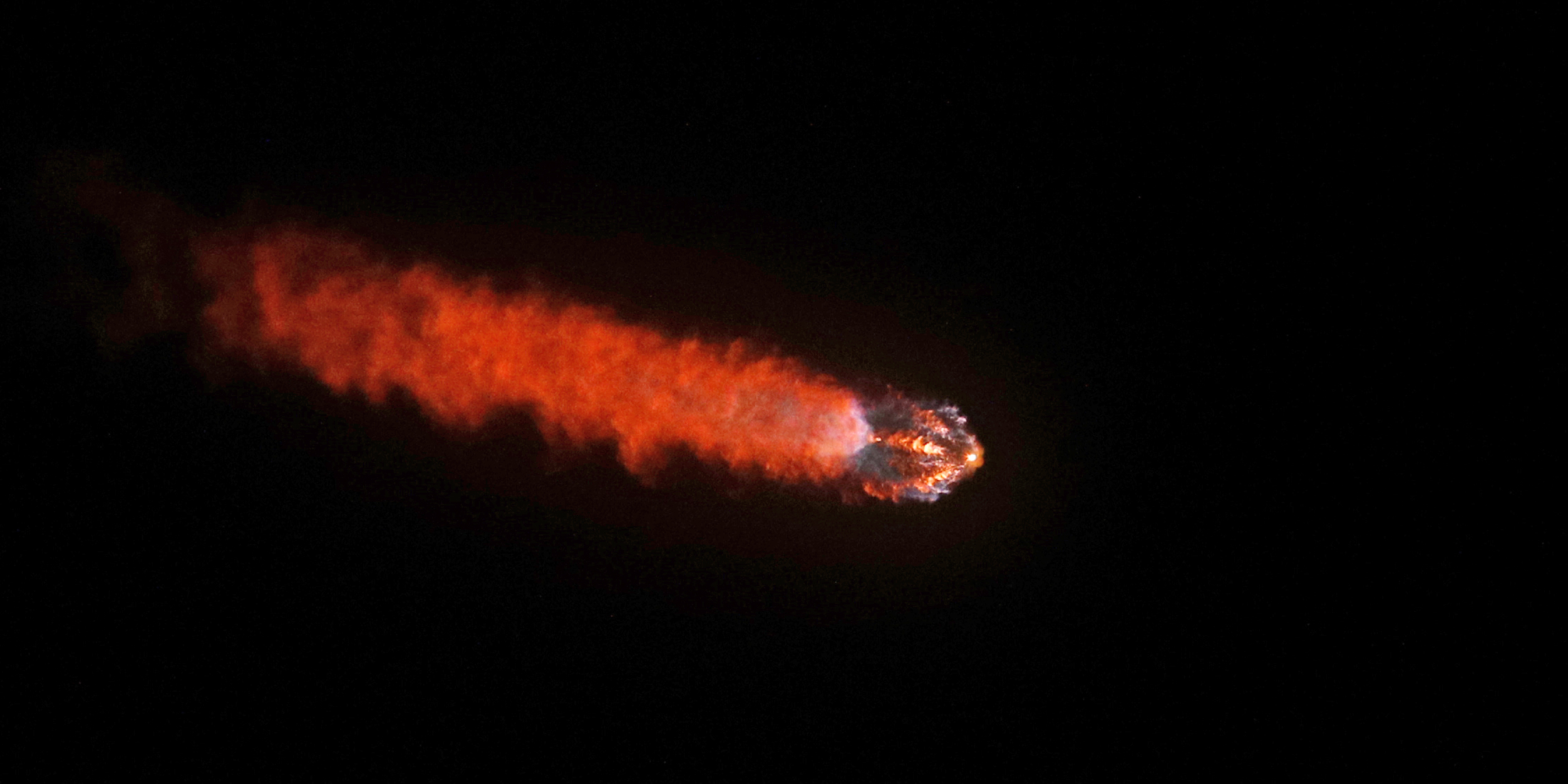
(Image: SpaceX Falcon 9 rocket for the Fram2 mission, by Gregg Newton / AFP via Getty Images)