On the 17th of December 2024, Glasgow Caledonian University hosted an engaging and educational taster session on X-Ray physics for S4 and S5 physics pupils from Dunoon Grammar. This event aimed to ignite students' interest in radiography and provide them with a hands-on experience that bridges classroom learning with real-world applications, as well as an excellent outreach and promotion opportunity for radiography.
The session kicked off with a warm welcome and an introduction to the profession. Pupils were given an overview of the career opportunities in both diagnostic and therapeutic radiography and the aims of the session. The pupils and teachers were excited as they settled in for a day of discovery and learning.
The next segment of the session was a lecture on the basics of X-Ray physics, delivered by Claire Currie, Senior Lecturer in Diagnostic Imaging. Pupils learned about the basic physical principles of X-rays, how they are produced, and some key concepts about interactions with the body and radiation safety. They were fascinated to learn that Roentgen discovered X-rays by accelerating electrons, well before J.J. Thomson discovered the electron! The lecture was interactive, with pupils engaging in a fun quiz. One of the questions that sparked interest was: "Can you tell me how X-rays are made? 1. Magic, 2. Harvested from the moon, 3. From X-ray seeds grown underground, 4. Electrons are accelerated and collide with a metal target." The correct answer, of course, is that electrons are accelerated and collide with a metal target.
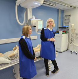
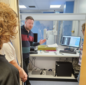
Following a short break, the pupils were given a safety briefing before visiting our X-Ray room. Emphasis was placed on the importance of radiation protection, personal dosimetry, and the use of personal protective shielding equipment. With safety protocols in place, the pupils eagerly participated in the practical session. The opportunity to visit the GCU X-ray room was ably supported and supervised by Will MacGregor and Sharon Stewart, Lecturers in Diagnostic Imaging. The pupils enjoyed demonstrations of the equipment and even tried on some lead protection, which added a fun and educational element to the experience.
In another room, Karen Cameron, Lecturer in Radiotherapy and Oncology, provided insights into the profession. The pupils were fascinated by the applications of radiotherapy and the technology involved.
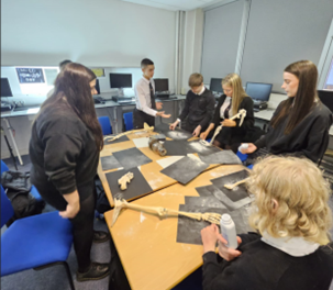
Back in the classroom, we set up an ‘X-ray museum’ with X-ray tubes, imaging receptor and other equipment parts. Pupils created their own radiographs using bones, talc, and black paper, which allowed them to put theory into practice in a creative and engaging way. After the hands-on experience, students gathered for a group discussion to share their findings and observations. This was followed by an interactive activity where they brainstormed real-life applications of X-rays created attractive posters on topics related to radiation protection to take back to their school.
Mr. McNeilage and the other science teachers expressed their thanks on social media, noting that the pupils were "raving about the trip the whole way home, and it was the most interactive trip they'd experienced." The feedback from the teachers highlighted the success of the session in providing an immersive and educational experience for their pupils.
As Radiography lecturers, we found this outreach project to be a fun and engaging way to promote the profession. We would be happy to engage with other Radiographers about this outreach project and share our resources.
Get in touch with [email protected]


Photographs shared with permission.
